 Add Row
  Add
Add 


Psittacine Beak and Feather Disease (PBFD) is an infectious, fatal viral illness that impacts the beak, plumes, and body immune system of birds coming from the Psittacidae family. It was very first recognized in 1975 by vets in Australia, where the illness affects wild birds. Although birds revealing signs of disease generally pass away, it is common for birds to be exposed to the infection, establish a mild infection, and recover.
What birds are at risk for PBFD?
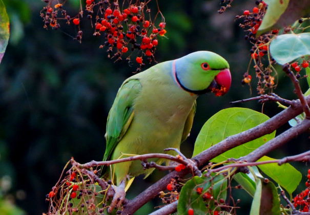
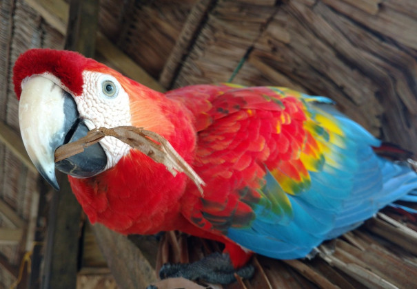
PBFD has actually been identified in over 40 species of psittacines, primarily in Old World members of the parrot family. PBFD is seen more frequently in cockatoos, however Eclectus parrots, lovebirds, budgies, and African grey parrots are likewise impacted. Younger birds are more commonly affected, particularly with the intense form of the illness. Most birds detected with psittacine beak and feather disease are under 2 years of age.
What triggers PBFD?
PBFD is caused by a DNA infection that affects the cells of the immune system and those that produce the beak and plumes.
The virus is a circovirus, which is among the smallest infections understood to cause illness. A comparable virus impacts doves and other birds.
How is the infection that causes PBFD transferred?
PBFD is extremely infectious. Big quantities of the infection, which can end up being airborne, are discovered in the droppings, contents of the crop, and the feather dust of contaminated birds. The plume dust is easily distributed and can contaminate food, water, cages, clothing, and other locations of the environment. Psittaccine beak and feather disease is thought to be transferred by inhalation or intake of the infection. It has been recommended that the virus might be transferred in utero from the female bird to the egg.
The incubation duration (time in between direct exposure to the virus and the advancement of indications) can be as short as 3-4 weeks, or up to several years, relying on the quantity of infection transmitted, the age of the bird, the phase of plume advancement, and the health of the bird's body immune system.

What are the signs of PBFD?
There are both intense and chronic forms of the disease.
PERACUTE/ACUTE FORM:
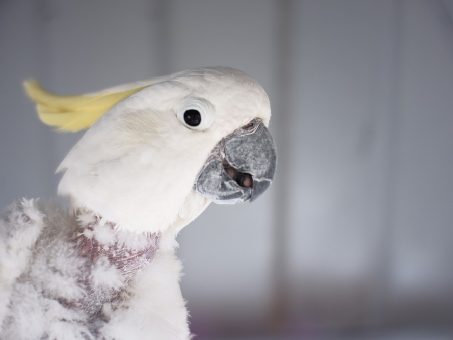
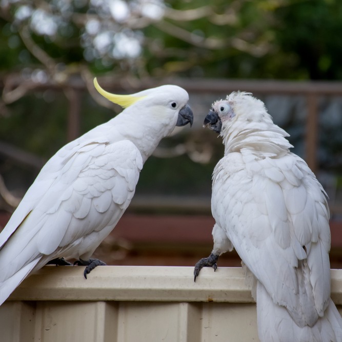
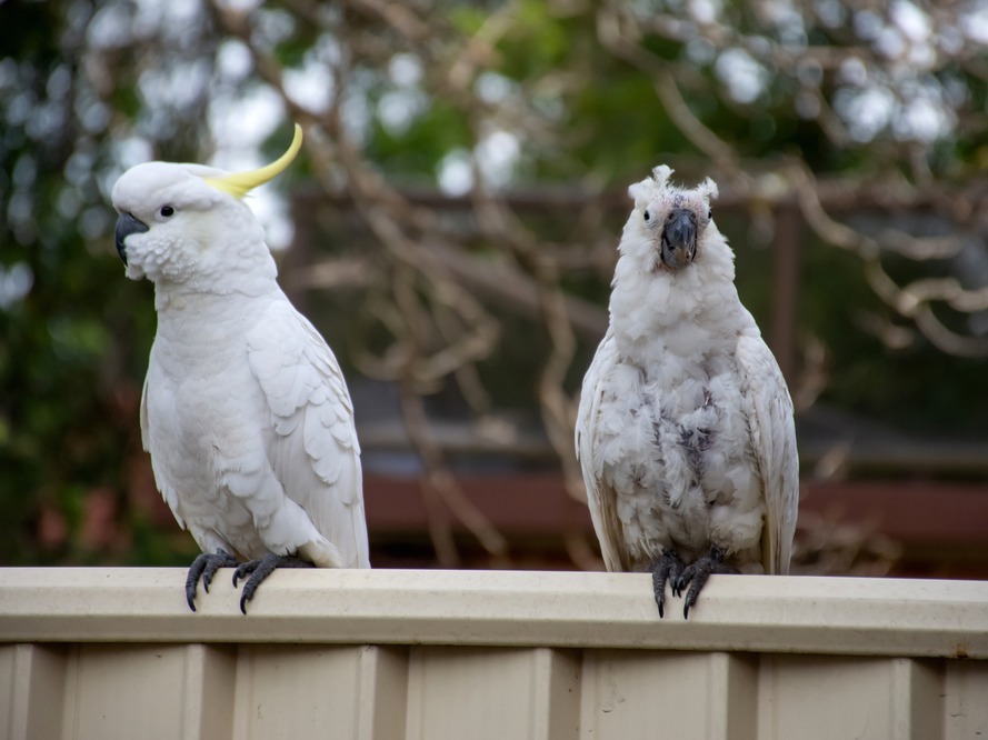
The peracute and acute types most commonly happen in extremely young birds, and might begin with signs unassociated to the beak or plumes. Affected birds are frequently depressed and throw up due to crop tension. They may establish a diarrhea-causing enteritis or pneumonia, and die without displaying any sores of the plumes or beak. This is typically called the peracute kind of the disease. In the intense kind, juveniles losing their down and establishing feathers may have lesions on the feathers, consisting of circular bands around the plumes which restrict the plume at its base. These feathers are often loose, break quickly, may bleed, and are extremely unpleasant.
Typical Signs of Psittacine Beak & & Feather Disease
Acute FormChronic FormDepressionLoss of plume dust and powderRegurgitation and diarrheaAbnormal feather developmentLoss of appetite and weightAbnormal growth and deformities of the beakAbnormal feather developmentNecrotic beak and oral lesionsDeathSecondary infectionsDeath in months to yearsCHRONIC FORM:
In the chronic kind of PBFD, which is more typical in older birds, the powder-down plumes are typically the first plumes affected.
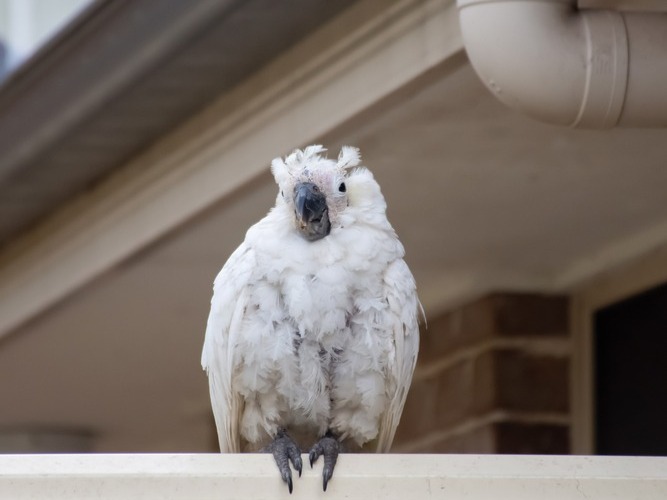
The plumes are fragile and fracture easily, have constricting bands, might hemorrhage, and may be blemished, deformed, or curled. As the plume roots are damaged, the bird will quickly be not able to replace feathers, and the main, secondary, tail, and crest feathers are lost. Bare skin is exposed, and the normal feather dust is not found on the body or the beak, where it generally builds up due to preening. Plume irregularities, often described "dystrophic plumes," might not appear till the first molt after infection, which could be a period approximately 6 months.
The beak might establish irregular sunken areas. Brown lethal areas might be discovered inside the upper beak, and the beak may extend, become deformed, and fracture. Secondary beak and oral infections often occur. In some birds, the nails can likewise be deformed or slough.
Mucus in the droppings, or a green tint to the droppings may occur.
In some birds, the liver will be impacted, and liver failure might be the cause of death.
Birds with the persistent form of the illness may live for months to years before passing away of a secondary infection. This extended period of health problem in which the bird might be featherless, and gradually deteriorates can be extremely emotionally hard for owners.
How is PBFD detected?
The review of the case history, existence of clinical indications, and observations throughout the physical examination support the medical diagnosis of PBFD. Other conditions such as nutritional shortages, infection with polyomavirus (causes budgerigar fledgling illness and other diseases of psittacines), hormonal problems, and drug responses can cause lesions on the plumes comparable to PBFD. Histopathology (tiny evaluations of biopsies) can confirm the medical diagnosis. Impacted cells will have irregularities in their nuclei, called "basophilic intranuclear addition bodies." The medical diagnosis may likewise be confirmed by a PCR (polymerase domino effect) test on entire blood or biopsy samples from the affected bird.

The test spots the presence of the virus. This test may likewise be used on swabs of surface areas in the environment to identify contamination.
Incorrect positive and false unfavorable test results can happen. For example, contaminated air-borne cells could contaminate a sample and cause a false positive result. Healthy birds with a favorable test result should be retested after 90 days. If they still have favorable test outcomes, they should be thought about providers of the infection. If the retest is unfavorable, the bird might have gotten rid of the infection, and become immune.
False negative outcomes may happen if too much anticoagulant exists in the sample, an incredibly high variety of viral particles exist and interfere with the test, or there are an insufficient number of infected leukocyte in the sample.
How is PBFD dealt with?
There is no particular treatment for PBFD. Supportive care consisting of great nutrition, supplemental heat (incubator), beak cutting, and treatment of secondary infections can be used.

The illness, nevertheless, is progressive, and very few birds recover. Euthanasia might need to be considered for birds with severe and/or unpleasant signs. Birds who pass away a natural death usually succumb to a secondary bacterial, fungal, or viral infection regardless of treatment, considering that their immune systems have actually been seriously reduced. A lot of birds die within 6 months to 2 years of establishing the illness.
How is PBFD avoided and managed?
Birds needs to be purchased from suppliers with disease-free birds. New birds entering into centers should be quarantined and tested. Repeat screening in 3-4 weeks to permit the incubation duration is advised. Infected birds ought to be separated and eliminated from breeding programs. Juvenile birds ought to be housed independently from grownups. Bird owners need to understand that if they manage other individuals' birds, it might be possible for them to bring the virus into their house and infect their birds.
Excellent hygiene and sanitation need to be utilized. There is no recognized disinfectant that kills this virus.
In Australia, a killed vaccine has actually been established which can protect unexposed birds; it can trigger more severe illness in birds currently revealing indications of PBFD. Birds must be vaccinated as young as possible, as soon as 14 days of age. The vaccine ought to be boostered after one month, and reproducing birds need to be vaccinated one month prior to reproducing.

Write A Comment